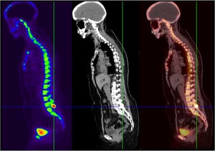
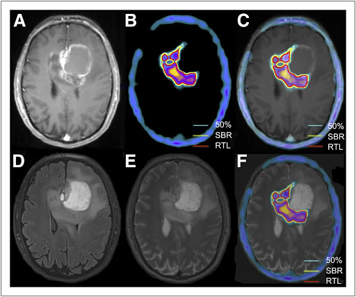
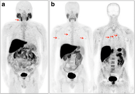
A positron emission tomography (PET) scan is an imaging method using positron emitting radionuclides labelled with different pharmaceuticals and studies their bio-distribution (Emission scan).This when combined with CT scan (transmission) scan is called as PET-CT scan
The tracer may be injected, swallowed or inhaled, depending on which organ or tissue is being studied by the PET scan. The tracer accumulates in areas of your body that have higher levels of chemical activity, which often correspond to areas of disease. On a PET scan, these areas show up as bright spots.
A PET scan is useful in detecting and staging/Grading various ailments like cancer, coronary artery disease, tuberculosis and other inflammations
PET –CT scan is an effective procedure, which assists in detecting chemical activity levels in the body. It is helpful in ruling out various disorders in brain and heart, including cancers. It is more effective than CT and conventional MRI has molecular changes appear earlier than morphological changes. There can be multiple conditions where PET- CT scan is very useful. Some of the disease conditions are as mentioned.
Cancerous lesions are often seen as bright spots in a PET-CT scan as the cancer cells have high levels of metabolic rate as compared to the normal cells. Thus the PET scans are helpful in the following scenarios:
It is very important to read the findings on a PET scan very carefully as some benign lesions show abnormal metabolic activity. There can be multiple types of cancers, which can be detected and diagnosed by PET scans. Some of these are highlighted below.
Multiple different types of heart related disorders can be diagnosed with PET scans. Some of these disorders include.

Fluorodeoxyglucose PET or FDG PET
Since tumours need sugars to grow, fluorodeoxyglucose is used. It is a sugar-like substance with the ability to bind with sugars. When it is introduced into the body, it gravitates towards with highconcentrations of sugar. While it can be used to find tumours in any part of the body, it is specifically useful to image inflammation, infection, and brain function.

DOTA-NOC PET
This drug is typically used to detect neural or neuroendocrine tumours.

Fluoride Bone PET
As the name suggests, it is useful to create images of bones.

Fluorothymidine PET or FLT-PET
FLT helps to detect fast-replicating cells and is typically used for imaging bone marrow, brain tumours, and other clinical trials.

Fluorocholine PET or FCH-PET
This drug is used to image prostate cancer
ONLY
After the procedure, the patient is asked to change into normal clothes and is advised to drink plenty of water/fluids in order to flush out or remove the tracer from the body. The patient can go home after the PET-CT scan is performed and resume normal routine activities. The patient can drink and eat normally after the PET –CT scan procedure is complete.
The nuclear physician interprets the images from the scan and prepares the report, which will further be handed over to the concerned doctor. The doctor examines the report carefully and discusses the results with the patient in detail. If the report is normal, it means the patient is fit and healthy with no abnormality detected. If the report is abnormal, it means that there are some abnormalities, which would need the attention of the doctor. The cancerous cells appear as bright spots in the image. The bright spots indicate cancer cells with high metabolic activity.
In order to diagnose and monitor cancer and its ongoing treatment, a PET CT Scan is prescribed. As many people are getting affected with cancer in India, PET CT Scans are required by medical doctors as they are a major key for cancer treatment.
Reasons for using PET Scan:
The following are the prerequisites for a PET CT scan if the contrast dye is used in the procedure.
A typical CT scan usually costs around ₹4000, which may go as high as ₹17,000 depending upon the area of the body to be scanned and also on the location of the medical institution offering the scan
During the process of a CT scan, you will be exposed to radiation that will be specifically targeted to a particular area. The impact of these radiations is negligible and hence, CT scans are not only completely safe but also the accuracy with which they provide the results helps protect people from uncountable life-threatening diseases.
A typical HRCT scan takes around 30-60 minutes. The majority of the time required for an HRCT scan goes into preparing the person for the HRCT scan, whereas the actual test is done within a few quick minutes
Whether you can eat or not depends upon the type of CT scan your doctor asked for. If diagnosis demands a CT scan with contrast, It is crucial that you don’t eat anything three hours before the appointment and if it demands a CT scan without contrast there are no restrictions on consumption of food, fluids, and medicines before the test.
The basic point of difference between a CT scan and an MRI scan is the radiation they use to produce the image of the scanned area. In the case of CT scan, the radiations used are X-rays which makes CT scans comparatively cheaper but the downside is that the image produced lacks details and only gives a rough idea in the case of MRI scan, magnetic and radio waves are used, the use of which makes this procedure costlier but on the brighter side, the image produced is highly detailed.
